Find out how NHSGGC Improves PICC Placement Safety with ECG Tip Location System pilotTLS

Glasgow hospital improves peripherally inserted central catheter (PICC) placement safety with Vygon’s pilotTLS™ ECG Tip Location System
Nov 2020
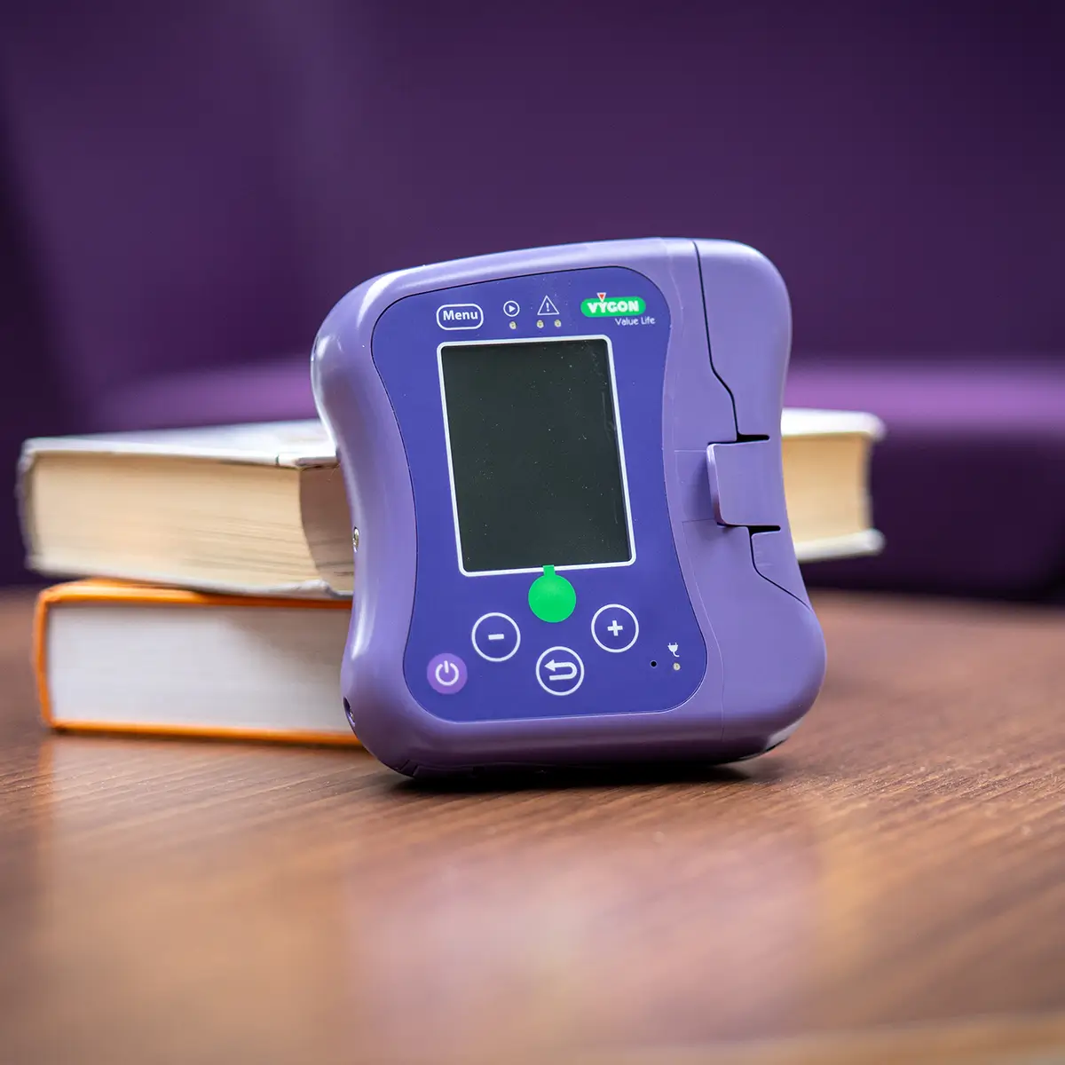
Introducing Vygon’s pilotTLS™ ECG Tip Location System to confirm the accurate placement of PICCs has improved both the safety and experience of patients at the Queen Elizabeth University Hospital in Glasgow – part of the NHS Greater Glasgow & Clyde (NHSGGC). Each year the demand on the Vascular Access Service (VAS) in NHSGGC has grown, with 3,213 PICC lines inserted in 2019. The precise placement of a PICC tip is important in order to ensure good catheter performance and to prevent a wide range of infusion-related complications. Prior to using pilotTLS™, a post-procedure chest x-ray was used to confirm the catheter tip position.
The evaluation of the pilotTLS™ ECG Tip Location System at the Queen Elizabeth University Hospital at the end of 2019 was led by Nicola Wyllie, the Senior Charge Nurse for the VAS.
Nicola was prompted to evaluate tip placement methods after undertaking a literature review of post-procedure chest x-ray and anatomical landmarks after PICC placement.
A European multicentre study by Pittiruti et al(1) confirmed that the intracavitary ECG method for real-time verification of tip position is accurate, safe, feasible in all adult patients, and applicable to any type of short-term or long-term central venous access device.
Nicola reviewed the new technologies available using the catheter as an intra-cavitary electrode. The pilotTLS™ focuses on the changes in the P wave of the PQRS complex allowing for greater accuracy of tip location than traditional methods.
Changing to the pilotTLS™ system means a post-procedure placement x-ray is no longer needed after PICCs are inserted. This is in line with Ionising Radiation Medical Exposure Regulations (IRMER) stating the need to use alternatives to x-rays where appropriate so that a patient’s exposure to ionising radiation is minimised. The use of ECG method would require less input from a radiologist or radiographer, allowing capacity improvement in that area. With fewer requirements to move patients from placement table to x-ray suite, there is also a reduction in the burden on porters and other staff involved in the movement of patients.
“We found the equipment very straightforward to use and we became confident very quickly,” explained Nicola. “There was no requirement for post procedure chest x-ray meaning the patient is with the service for a shorter time. In fact, it has improved the efficiency of the imaging department as a whole with less patients waiting for the x-ray”.
As well as the safety benefits, using the pilotTLS™ is a quicker, simpler procedure and less traumatic for patients. Tip location can be done in real-time so, if the catheter is not placed correctly, it can be re-positioned immediately.
“Using anatomic land marks on a post procedure chest x-ray is not accurate, whilst using pilotTLS™ allows us to place the catheter at the optimal position every time. Real time indication of malpositioned catheters allows the VAS to manipulate the catheter at time of insertion rather that following confirmation on x-ray. In addition, the system is portable, so PICCs can be accessed immediately following a bedside placement with no requirement to wait for portable chest x-ray”.
During the evaluation process, the VAS team continued to use post-procedure chest x-ray for four weeks alongside the pilotTLS™ to ensure they were using the equipment appropriately.
A total of 72 PICCs were placed during the evaluation period and the pilotTLS™ was used on 59 of the patients. Ten were inserted in the department whilst the pilotTLS™ was being used at another patient’s bedside, and the remaining three patients had pacemakers in situ.
The costs of the two procedures were also assessed. Consumables for the pilotTLS™ were £16.72 per procedure compared to x-ray costs of around £65 per procedure (excluding subsequent attempts for malpositioned tips). This means the hospital can realise savings of around £48.73 per procedure, equating to £156,569 per annum based on their 2019 demand of 3,213 PICCs. Nicola also identified further benefits of the pilotTLS™ system compared to alternatives. She added:
“We receive a large number of referrals from cardiology and the AF mode on the pilotTLS™ system allowed us to use the equipment on this patient group. To my knowledge this is the only system with this function.” “The use of the pilotTLS™ has improved the experience of our patients and the efficiency of our service”
References
1. Pittiruti M, Bertollo D, Briglia E, et al. The intracavitary ECG method for positioning the tip of central venous catheters: results of an Italian multicenter study. J Vasc Access. 2012;13(3):357–365
Learn more about how pilotTLS can help you
To speak to one of our expert Sales Managers please complete the below form.


